


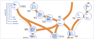


 Foundational characteristics of cancer include proliferation, angiogenesis, migration, evasion of apoptosis, and cellular immortality. Find key markers for these cellular processes and antibodies to detect them.
 The SUMOplot™ Analysis Program predicts and scores sumoylation sites in your protein. SUMOylation is a post-translational modification involved in various cellular processes, such as nuclear-cytosolic transport, transcriptional regulation, apoptosis, protein stability, response to stress, and progression through the cell cycle.
 The Autophagy Receptor Motif Plotter predicts and scores autophagy receptor binding sites in your protein. Identifying proteins connected to this pathway is critical to understanding the role of autophagy in physiological as well as pathological processes such as development, differentiation, neurodegenerative diseases, stress, infection, and cancer.
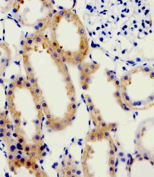
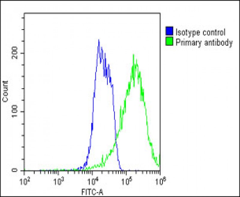
ADRA1D Antibody (N-term)
Purified Rabbit Polyclonal Antibody (Pab)
- SPECIFICATION
- CITATIONS: 1
- PROTOCOLS
- BACKGROUND

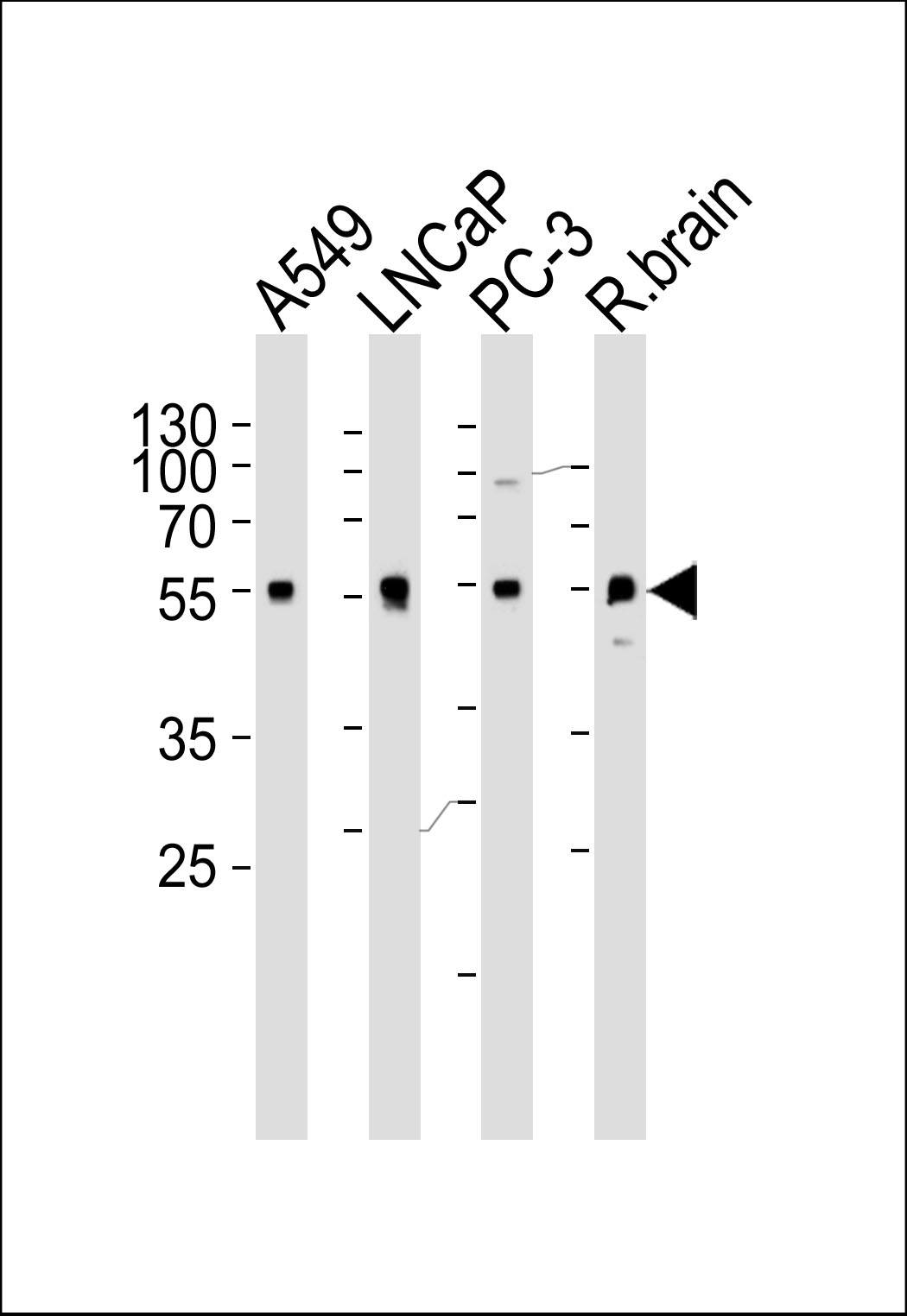
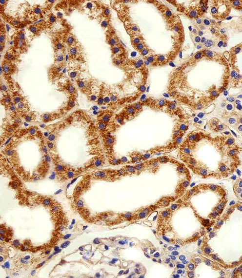
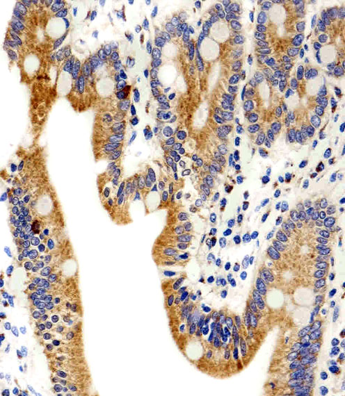
Application
| WB, IHC-P, FC, E |
|---|---|
| Primary Accession | P25100 |
| Reactivity | Human, Rat |
| Predicted | Mouse, Rabbit, Bovine, Dog, Sheep |
| Host | Rabbit |
| Clonality | Polyclonal |
| Isotype | Rabbit IgG |
| Calculated MW | 60463 Da |
| Antigen Region | 1-30 aa |
| Gene ID | 146 |
|---|---|
| Other Names | Alpha-1D adrenergic receptor, Alpha-1A adrenergic receptor, Alpha-1D adrenoreceptor, Alpha-1D adrenoceptor, Alpha-adrenergic receptor 1a, ADRA1D, ADRA1A |
| Target/Specificity | This ADRA1D antibody is generated from a rabbit immunized with a KLH conjugated synthetic peptide between 1-30amino acids from the N-terminal region of human ADRA1D. |
| Dilution | WB~~1:1000 IHC-P~~1:25 FC~~1:25 |
| Format | Purified polyclonal antibody supplied in PBS with 0.09% (W/V) sodium azide. This antibody is purified through a protein A column, followed by peptide affinity purification. |
| Storage | Maintain refrigerated at 2-8°C for up to 2 weeks. For long term storage store at -20°C in small aliquots to prevent freeze-thaw cycles. |
| Precautions | ADRA1D Antibody (N-term) is for research use only and not for use in diagnostic or therapeutic procedures. |
| Name | ADRA1D |
|---|---|
| Synonyms | ADRA1A |
| Function | This alpha-adrenergic receptor mediates its effect through the influx of extracellular calcium. |
| Cellular Location | Cell membrane; Multi-pass membrane protein. |

Provided below are standard protocols that you may find useful for product applications.
Background
This alpha-adrenergic receptor mediates its effect through the influx of extracellular calcium.
References
Bruno J.F.,et al.Biochem. Biophys. Res. Commun. 179:1485-1490(1991).
Forray C.,et al.Mol. Pharmacol. 45:703-708(1994).
Schwinn D.A.,et al.J. Pharmacol. Exp. Ther. 272:134-142(1995).
Weinberg D.H.,et al.Biochem. Biophys. Res. Commun. 201:1296-1304(1994).
Esbenshade T.A.,et al.Mol. Pharmacol. 47:977-985(1995).
If you have used an Abcepta product and would like to share how it has performed, please click on the "Submit Review" button and provide the requested information. Our staff will examine and post your review and contact you if needed.
If you have any additional inquiries please email technical services at tech@abcepta.com.
Ordering Information
Other Products
Shipping Information