


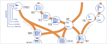


 Foundational characteristics of cancer include proliferation, angiogenesis, migration, evasion of apoptosis, and cellular immortality. Find key markers for these cellular processes and antibodies to detect them.
 The SUMOplot™ Analysis Program predicts and scores sumoylation sites in your protein. SUMOylation is a post-translational modification involved in various cellular processes, such as nuclear-cytosolic transport, transcriptional regulation, apoptosis, protein stability, response to stress, and progression through the cell cycle.
 The Autophagy Receptor Motif Plotter predicts and scores autophagy receptor binding sites in your protein. Identifying proteins connected to this pathway is critical to understanding the role of autophagy in physiological as well as pathological processes such as development, differentiation, neurodegenerative diseases, stress, infection, and cancer.
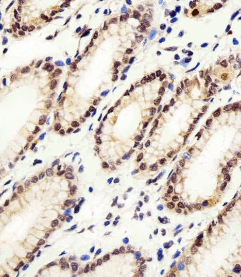
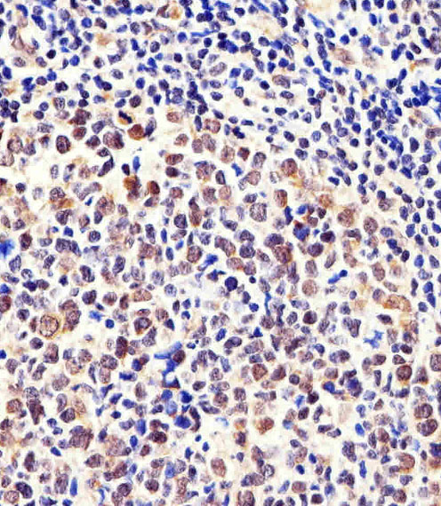
Cellular Apoptosis Susceptibility Antibody (C-term)
Purified Rabbit Polyclonal Antibody (Pab)
- SPECIFICATION
- CITATIONS: 3
- PROTOCOLS
- BACKGROUND

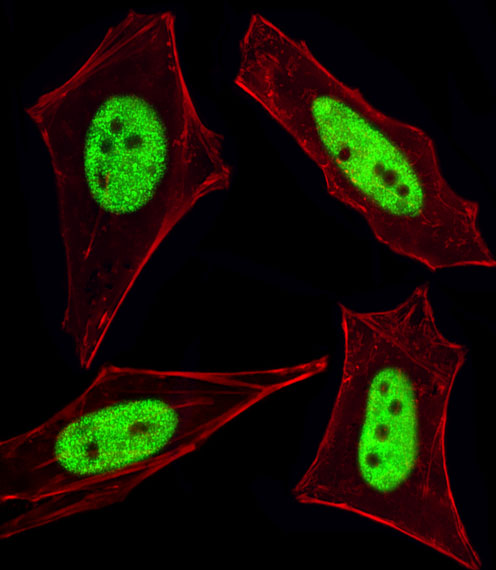
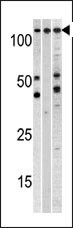
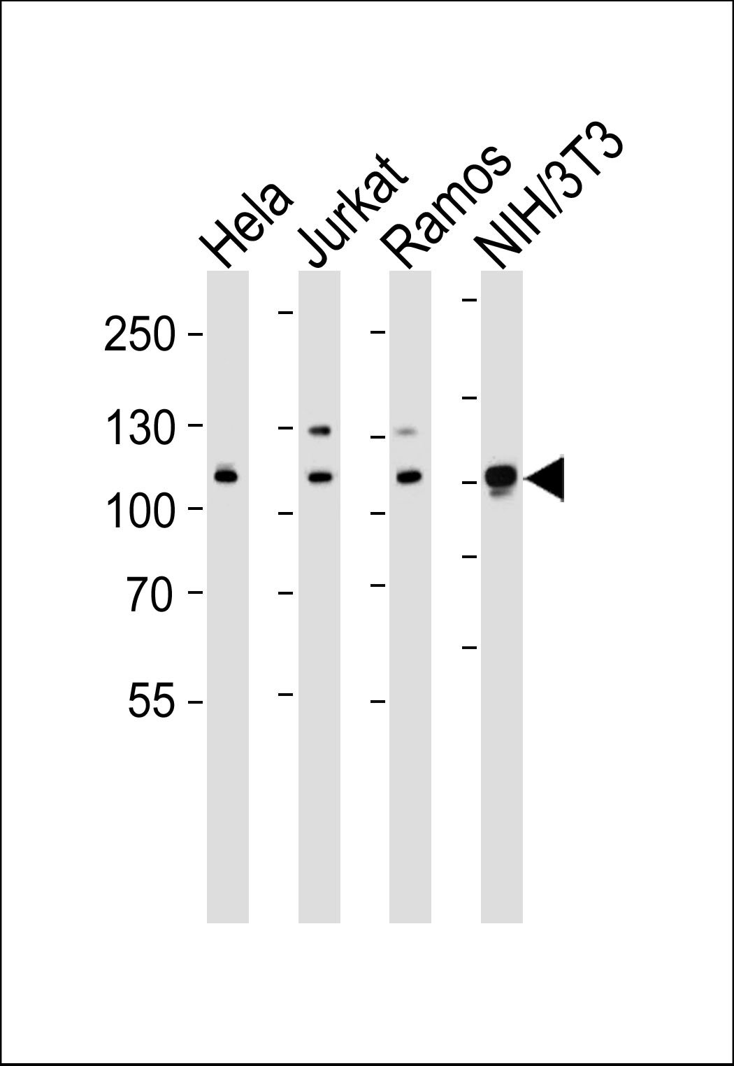
Application
| IHC-P, IF, WB, E |
|---|---|
| Primary Accession | P55060 |
| Other Accession | Q9ERK4, Q7SZC2, A5D785 |
| Reactivity | Human, Mouse |
| Predicted | Bovine, Zebrafish |
| Host | Rabbit |
| Clonality | Polyclonal |
| Isotype | Rabbit IgG |
| Calculated MW | 110417 Da |
| Antigen Region | 55-84 aa |
| Gene ID | 1434 |
|---|---|
| Other Names | Exportin-2, Exp2, Cellular apoptosis susceptibility protein, Chromosome segregation 1-like protein, Importin-alpha re-exporter, CSE1L, CAS, XPO2 |
| Target/Specificity | This Cellular Apoptosis Susceptibility antibody is generated from rabbits immunized with a KLH conjugated synthetic peptide between 55-84 amino acids from the N-terminal region of human Cellular Apoptosis Susceptibility. |
| Dilution | IF~~1:100 WB~~1:1000 IHC-P~~1:100 |
| Format | Purified polyclonal antibody supplied in PBS with 0.09% (W/V) sodium azide. This antibody is prepared by Saturated Ammonium Sulfate (SAS) precipitation followed by dialysis against PBS. |
| Storage | Maintain refrigerated at 2-8°C for up to 2 weeks. For long term storage store at -20°C in small aliquots to prevent freeze-thaw cycles. |
| Precautions | Cellular Apoptosis Susceptibility Antibody (C-term) is for research use only and not for use in diagnostic or therapeutic procedures. |
| Name | CSE1L |
|---|---|
| Synonyms | CAS {ECO:0000303|PubMed:7479798}, XPO2 |
| Function | Export receptor for importin-alpha. Mediates importin-alpha re-export from the nucleus to the cytoplasm after import substrates (cargos) have been released into the nucleoplasm. In the nucleus binds cooperatively to importin-alpha and to the GTPase Ran in its active GTP-bound form. Docking of this trimeric complex to the nuclear pore complex (NPC) is mediated through binding to nucleoporins. Upon transit of a nuclear export complex into the cytoplasm, disassembling of the complex and hydrolysis of Ran-GTP to Ran-GDP (induced by RANBP1 and RANGAP1, respectively) cause release of the importin-alpha from the export receptor. CSE1L/XPO2 then return to the nuclear compartment and mediate another round of transport. The directionality of nuclear export is thought to be conferred by an asymmetric distribution of the GTP- and GDP-bound forms of Ran between the cytoplasm and nucleus. |
| Cellular Location | Cytoplasm. Nucleus. Note=Shuttles between the nucleus and the cytoplasm. |
| Tissue Location | Detected in brain, placenta, ovary, testis and trachea (at protein level) (PubMed:10331944). Widely expressed (PubMed:10331944). Highly expressed in testis and in proliferating cells (PubMed:7479798, PubMed:10331944). |

Provided below are standard protocols that you may find useful for product applications.
Background
Proteins that carry a nuclear localization signal (NLS) are transported into the nucleus by the importin-alpha/beta heterodimer. Importin-alpha binds the NLS, while importin-beta mediates translocation through the nuclear pore complex. After translocation, RanGTP binds importin-beta and displaces importin-alpha. Importin-alpha must then be returned to the cytoplasm, leaving the NLS protein behind. CSE1L binds strongly to NLS-free importin-alpha, and this binding is released in the cytoplasm by the combined action of RANBP1 and RANGAP1. In addition, CSE1L may play a role both in apoptosis and in cell proliferation.
References
Goldberg, G.S., et al., J. Biol. Chem. 278(47):46533-46540 (2003).
Behrens, P., et al., Apoptosis 8(1):39-44 (2003).
Jiang, M.C., et al., Biochem. Biophys. Res. Commun. 294(4):900-905 (2002).
Wellmann, A., et al., Int. J. Mol. Med. 7(5):489-494 (2001).
Brinkmann, U., et al., Genomics 58(1):41-49 (1999).
If you have used an Abcepta product and would like to share how it has performed, please click on the "Submit Review" button and provide the requested information. Our staff will examine and post your review and contact you if needed.
If you have any additional inquiries please email technical services at tech@abcepta.com.
Ordering Information
Other Products
Shipping Information